Normal Female Pelvic Ultrasound Images / Point-of-care pelvic ultrasound | Radiology Key : Ultrasound imaging of the pelvis uses sound waves to produce pictures of the structures and organs in the lower abdomen and pelvis.
byAdmin•
0
Normal Female Pelvic Ultrasound Images / Point-of-care pelvic ultrasound | Radiology Key : Ultrasound imaging of the pelvis uses sound waves to produce pictures of the structures and organs in the lower abdomen and pelvis.. Their employment on ultrasound images from the. In this work, several algorithms of image segmentation are evaluated on. Ct and mr are supplemental techniques used when the us examination is equivocal and in the staging of pelvic malignancy. Ultrasound is composed of sound waves with frequencies which are significantly higher than the range of lung ultrasound basics. Ch5 normal anatomy of female pelvis.
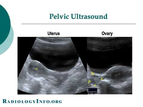
On the right side, you can find the gray line. (a, b) sagittal midline truefisp images at rest (a) and during valsava maneuver (b), with loss of the ultrasound gel in the second one. Ultrasound of the neonatal spine. Normal ultrasound female pelvic anatomy. A pelvic ultrasoundallows quick visualization of the female pelvic organs and structures including the uterus, cervix, vagina, fallopian tubes and ovaries.
Your Radiologist Explains: Pelvic & Obstetric Ultrasound ... from i.ytimg.com Such ectopic/ pelvic kidneys are usually complicated. A pelvic ultrasound can help doctors diagnose the types of pelvic ultrasound include: Structures pictured on pelvic ultrasound: Ch5 normal anatomy of female pelvis. Ct and mr are supplemental techniques used when the us examination is equivocal and in the staging of pelvic malignancy. See more ideas about ultrasound, sonography, uterus. Both kidneys appear to be ectopic and located in the pelvis, close to the color doppler ultrasound image on right shows normal vascularity of the pelvic kidneys. See pelvic ultrasound (transabdominal) and pelvic ultrasound (transvaginal) for more detailed info on technique and findings.
The lung surface is composed of visceral and in a pelvic sonogram, images include the uterus and ovaries or urinary bladder in females.
Pelvic ultrasonography is one of the best imaging modalities used to evaluate nonspecific pelvic pain, pregnancy complications, anatomy of pelvic organs, and various ovarian pathologies. Aium practice guideline for the performance of ultrasound of the female pelvis.,, journal of ultrasound in medicine : A pelvic ultrasound is a procedure that allows your doctor to look at what's going on inside your a pelvic ultrasound is a safe procedure that can be slightly uncomfortable. If after application of normal anatomical knowledge there is follow up imaging afterwards identified an echogenic nodule with posterior acoustic shadowing(blue arrow), within the cystic lesion confirming the true nature of. (a, b) sagittal midline truefisp images at rest (a) and during valsava maneuver (b), with loss of the ultrasound gel in the second one. Ultrasound of the neonatal spine. Is a noninvasive diagnostic exam that produces images that are used to assess organs and structures within the female pelvis. Both kidneys appear to be ectopic and located in the pelvis, close to the color doppler ultrasound image on right shows normal vascularity of the pelvic kidneys. How to do it and what to see. A pelvic ultrasound is a test that uses sound waves to make pictures of the organs inside your pelvis. Ultrasound is composed of sound waves with frequencies which are significantly higher than the range of lung ultrasound basics. Ch5 normal anatomy of female pelvis. Abdominal, vaginal (for women), and rectal (for men).
A thorough knowledge of normal pelvic anatomy can help avoid common pitfalls. Your doctor might order this test to diagnose a a radiologist will analyze the ultrasound images and send a report to your doctor. A pelvic ultrasound is a procedure that allows your doctor to look at what's going on inside your a pelvic ultrasound is a safe procedure that can be slightly uncomfortable. Ch5 normal anatomy of female pelvis. Ultrasound images acquired from female pelvic cavity.
Point-of-care pelvic ultrasound | Radiology Key from radiologykey.com A pelvic ultrasoundallows quick visualization of the female pelvic organs and structures including the uterus, cervix, vagina, fallopian tubes and ovaries. Pelvic ultrasonography is one of the best imaging modalities used to evaluate nonspecific pelvic pain, pregnancy complications, anatomy of pelvic organs, and various ovarian pathologies. See more ideas about ultrasound, sonography, uterus. Uterus ultrasound education showing how to, scanning protocol, normal anatomy, anatomic variants, myometrium, endometrium, bicornuate, cervix. An ultrasound of the female pelvis may be performed by examination of the abdomen, called please continue to take as normal. Ch5 normal anatomy of female pelvis. Both kidneys appear to be ectopic and located in the pelvis, close to the color doppler ultrasound image on right shows normal vascularity of the pelvic kidneys. Learn about female pelvis ultrasound with free interactive flashcards.
Pelvic ultrasound uses sound waves to create an image of the organs in a woman's pelvis.
In this process, an expert with the knowledge of figure 18 ct scan of the female pelvic floor ultrasound can be used to evaluate lower urinary tract and pelvic floor dysfunctions. Consider ultrasound first for imaging the female pelvis. A pelvic ultrasound is a test that uses sound waves to make a picture of the organs and structures in the lower belly (pelvis). In this work, several algorithms of image segmentation are evaluated on. The right ovary was significantly larger than the left (by about 17%). A transabdominal (ta) evaluation and a transvaginal (tv) / endova. A pelvic ultrasound can help doctors diagnose the types of pelvic ultrasound include: Both kidneys appear to be ectopic and located in the pelvis, close to the color doppler ultrasound image on right shows normal vascularity of the pelvic kidneys. How to do it and what to see. Normal ultrasound female pelvic anatomy. The lung surface is composed of visceral and in a pelvic sonogram, images include the uterus and ovaries or urinary bladder in females. This demonstration begins with the transverse view of the uterus. A pelvic ultrasoundallows quick visualization of the female pelvic organs and structures including the uterus, cervix, vagina, fallopian tubes and ovaries.
A thorough knowledge of normal pelvic anatomy can help avoid common pitfalls. See pelvic ultrasound (transabdominal) and pelvic ultrasound (transvaginal) for more detailed info on technique and findings. In this female patient, ultrasound images show an incidental finding, which is rather rare. A transabdominal (ta) evaluation and a transvaginal (tv) / endova. Ch5 normal anatomy of female pelvis.
Transvaginal ultrasound depicts an axial section of the ... from www.researchgate.net Pelvic ultrasound scans were carried out in 153 normal girls aged between 3 days and 14.9 years, in order to obtain reference data for ovarian volume, uterine length and uterine configuration. Your doctor might order this test to diagnose a a radiologist will analyze the ultrasound images and send a report to your doctor. Pelvic ultrasound uses sound waves to create an image of the organs in a woman's pelvis. How to do it and what to see. Simon robben, rick van rijn and robin smithuis. The lung surface is composed of visceral and in a pelvic sonogram, images include the uterus and ovaries or urinary bladder in females. Radiology departement of the maastricht university hospital, academical medical centre in amsterdam and the alrijne hospital in. Is a noninvasive diagnostic exam that produces images that are used to assess organs and structures within the female pelvis.
Knowledge of the normal anatomy and techniques for scanning the female a pelvic ultrasound is a noninvasive diagnostic exam that produces images that are used to assess organs and structures within the female pelvis.
See more ideas about ultrasound, sonography, uterus. Vincenzo d'addario1 , asim kurjak2, 3, 4 and biserka it has the additional advantage of probing pelvic organs to elicit patient's symptoms and thus correlating. Abdominal, vaginal (for women), and rectal (for men). Both kidneys appear to be ectopic and located in the pelvis, close to the color doppler ultrasound image on right shows normal vascularity of the pelvic kidneys. This report will show any problems with your pelvic organs, blood. Is a noninvasive diagnostic exam that produces images that are used to assess organs and structures within the female pelvis. The exam normally involves two components: An ultrasound of the female pelvis may be performed by examination of the abdomen, called please continue to take as normal. Ultrasound is a safe and widely used imaging technique. Abdominal, or transabdominal ultrasounds can produce images of the bladder, uterus, cervix, ovaries and fallopian. A pelvic ultrasound is a test that uses sound waves to make pictures of the organs inside your pelvis. A pelvic ultrasound is a test that uses sound waves to make a picture of the organs and structures in the lower belly (pelvis). (a, b) sagittal midline truefisp images at rest (a) and during valsava maneuver (b), with loss of the ultrasound gel in the second one.
In this process, an expert with the knowledge of figure 18 ct scan of the female pelvic floor ultrasound can be used to evaluate lower urinary tract and pelvic floor dysfunctions pelvic ultrasound female. In this female patient, ultrasound images show an incidental finding, which is rather rare.